Virology
Detection, Identification and Diagnosis of viruses
Serological / Immunological Methods
1) Haemagglutination
2) Haemagglutionation inhibition
3) Western Blot
4) D.ELISA
2) Haemagglutionation inhibition
3) Western Blot
4) D.ELISA
Haemagglutination
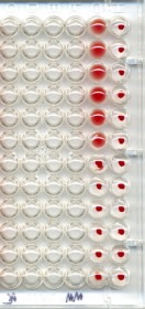
The Hemagglutination Assay(HA) is a quantification of viruses by hemagglutination. Some viral families have surface or envelope proteins, it is able to agglutinate(stick on to) human or animal Red blood cells (RBC) and binds to its N-acetylneuraminic acid. The method is simple and fast and may be used on large amounts of samples at the same time.
Haemagglutionation inhibition
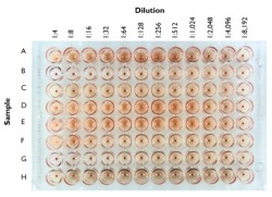
The basis of the hemagglutination inhibition(HI) assay is that antibodies to influenza virus will prevent attachment of the virus to red blood cells. Therefore hemagglutination is inhibited when antibodies are present. The highest dilution of serum that prevents hemagglutination is called the HI titer of the serum. If the serum contains no antibodies that react with the new H1N1 strain, then hemagglutination will be observed in all wells. Likewise, if antibodies to the virus are present, hemagglutination will not be observed until the antibodies are sufficiently diluted.
Western Blot

The western blot (protein immunoblot) is an analytical technique used to detect specific proteins in the given sample of tissue homogenate or extract. It uses gel electrophoresis to separate native or denatured proteins by the length or the polypeptide or by the 3-D structure of the protein. The proteins are then transferred to a membrane, where they are probed used antibodies specific to the target protein.
D.ELISA
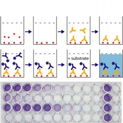
Enzyme-linked immunosorbent assay, also called ELISA. It is a biochemical technique used mainly in immunology to detect the presence of an antibody or an antigen in a sample. The ELISA has been used as a diagnostic tool in medicaine and plat pathology, even quality control check in some industries. In simple terms, in ELISA an unknown amount of antigen is affixed to a surface, and then a specific antibody is washed over the surface so that it can bind to the antigen. This antibody is linked to an enzyme, and in the final step a substance is added that the enzyme can convert to some detectable signal. Thus in the case of fluorescence ELISA, when light of the appropriate wavelength is shone upon the sample, any antigen/antibody complexes will fluoresce.
