Virology
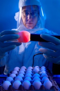
1. Embryonated eggs
Chick Embryo
- Embryonated hen's eggs are inoculated with animal viruses for the purpose of identification,
isolation, titration and for quantity cultivation in the production of viruses.
- Convenient, inexpensive host for many animal species
- If any viral growth occurs, it will be signaled by death of embryo/ embryo cell damage/
formation of typical pock (lesions) on membranes
- It is still used to grow viruses for some vaccines (eg. Influenza)
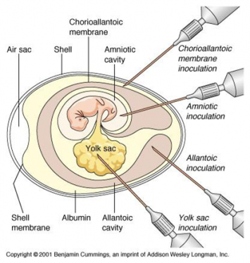
Illustration of sites of embryonated egg inoculation
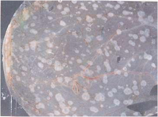
Pocks/Lesions
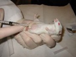
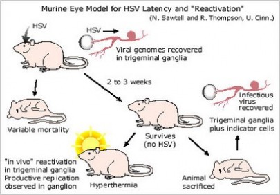
2. Animals
Why animals are use for research?
Medical researchers need to understand health problems before they can develop ways to treat them. Some diseases and health problems involve processes that can only be studied in a living organism. Animals are necessary to medical research when it is impractical or unethical to use humans.
Animals make good research subjects for a variety of reasons. Animals are biologically similar to humans. They are susceptible to many of the same health problems, and they have short life-cycles so they can easily be studied throughout their whole life-span or across several generations. In addition, scientists can easily control the environment around the animal (diet, temperature, lighting, etc.), which would be difficult to do with people. However, the most important reason why animals are used is that it would be wrong to deliberately expose human beings to health risks in order to observe the course of a disease.
Animals make good research subjects for a variety of reasons. Animals are biologically similar to humans. They are susceptible to many of the same health problems, and they have short life-cycles so they can easily be studied throughout their whole life-span or across several generations. In addition, scientists can easily control the environment around the animal (diet, temperature, lighting, etc.), which would be difficult to do with people. However, the most important reason why animals are used is that it would be wrong to deliberately expose human beings to health risks in order to observe the course of a disease.
Features
- Animal should be healthy and free from any dieseases
- Site of administration varies with the type of virus allowed to grow in the animal
- Animals slaughtered to harvest viruses
- Virally infected live animals are used to study the human body's immune system response to
viral infections
- This method is costly but still used with some exotic virus
- Site of administration varies with the type of virus allowed to grow in the animal
- Animals slaughtered to harvest viruses
- Virally infected live animals are used to study the human body's immune system response to
viral infections
- This method is costly but still used with some exotic virus
An example of using animals for studying viruses
Our understanding of the retrovirus that causes HIV/AIDS comes in part from studies of similar viruses in chickens, cats, and monkeys. Promising drugs and possible vaccines are tested first in mice and monkeys before being used in clinical trials with human volunteers.

Monkeys
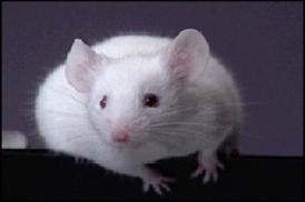
Mouse
Mouse infection
Mouse infection
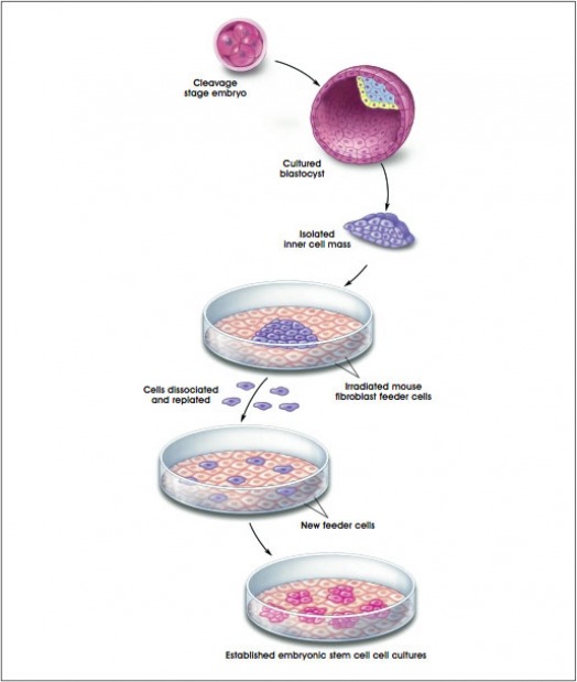
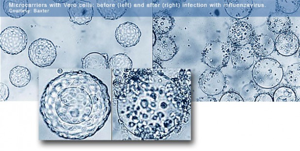
3. Cell Culture/Tissue Culture
- Cell culture is the process by which cells are grown under controlled conditions.
- Cells are grown in vitro on glass or a treated plastic surface in a suitable growth medium.
- In vitro: Literally in glass, as in a test tube. A test that is performed in vitro is one that is done
in glass or plastic vessels in the laboratory.
- It is a preferred method over embryonated eggs as it is more convenient and easier to
handle. However, some viruses cannot be grown in vitro
Primary cell line
Primary cell line are tissue cells that grow for only a few generations in vitro.
eg. Mouse primary cell line and Chick primary cell line.
eg. Mouse primary cell line and Chick primary cell line.
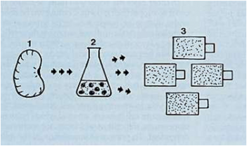
Piece of tissue (eg. Monkey kidney) used to produce tissue culture
Trypsinization
For experimental purposes, cells are often cultivated in containers that take the form of plastic flasks or plates. In such flasks, cells are provided with growth medium comprising the essential nutrients required for proliferation, and the cells adhere to the container and each other as they grow.
This process of cell culture requires a method to dissociate the cells from the container and each other. Trypsin, an enzyme can be used to "digest" the proteins that facilitate adhesion to the container and between cells.
The process of trypsinization is often done to permit passaging the cells to a new container, observation for experimentation, or reduction of the degree of confluency in the flask by removal of a percentage of the cells.
Continuous cell line
Continuous cell lines are derived from primary cell lines.They are transformed /cancerous cells. These cells are altered by effective dose of UV light and natural selection pressure yields cells that can grow well under in vitro conditions. They can be sub-cultured indefinitely and exist in either polyploid form or multiploid form. It is a method of choice to cultivate virus, where possible.
Continuous cell lines (cancerous cells present)
Methods for Viral Growth
1. Cytopathic Effect (CPE)
Cytopathic effect (CPE) refers to deterioration in cells (especially in tissue
culture) associated with the multiplication of certain viruses. When in tissue culture, the
spread of virus is restricted by an overlay of agar (or other suitable substance) and thus the
cytopathic effect may lead to formation of plaque.
The changes in appearance depending on the particular virus and type of cell
culture includes:
- Altered Shape
- Detachment from substrate
- Lysis
- Membrane fusion
- Altered membrane permeability
- Inclusion bodies
- Apoptosis (process of programmed cell death that may occur in multicellular organisms)
Cytopathic effects of Kunjin virus on Vero cells
The cytopathic effects of Kunjin virus on the Vero cells are that difference in appearance of the infected Vero cells as compared to the healthy Vero cells are observed. Most of the infected Vero cells which is a host to the Kunjin virus appeared to have merged together. As a result, most of the Vero cells cease growing in vitro when they come close to another cell, a phenomenon known as contact inhibition. Viruses are capable of causing cancer transformation. These transformation results in abnormal cell shape that does not recognize contact inhibition. The loss of contact inhibition results in unregulated growth. Hence as compared to the healthy Vero cells, the infected Vero cells appear to be more irregular in shape.
The cytoplasm of the infected Vero cells also contains numerous vacuoles with the Kunjin virus in it. The inclusion bodies are granules found in the cytoplasm or nucleus of some infected cells. These granules are viral parts. Dead cells can also be observed floating in the cell culture where the nucleus in the cells are absent. All in all, the cells become more separated and rounded as they deteriorate.
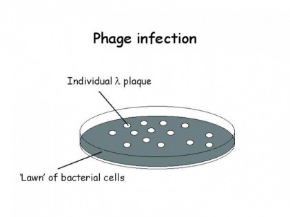
2. Plaque Assay
Direct counting is the actual counting of microorganisms. The basis of direct count is the actual counting of every organism present in a sub-sample of a population. However, microorganisms such as viruses are too small to be enumerated by direct counting. In order to enumerate viruses, many indirect methods have been developed. The most basic, simple and commonly used method is the plaque assay.
The theory of plaque assay is based on the original plaque assay for bacteriophages. Plaque assay helps to determine the quantity of infectious virus. This method requires the diluted virus to infect a monolayer cell line. The diluted virus is obtained by conducting serial dilution of the given virus. The concentration of the diluted virus is low hence one virus infects one cell and spreads to the surrounding cells. The infected monolayer is then grown under a gel medium (i.e. gelling medium like molten agar), after a virus burst only adjacent cells can be infected resulting in the formation of a visible circular plaque which indicate cell death as more cells are infected.
Principles of plaque assay
1. The concentration of virus is low enough that 1 virus infects 1 cell.
2. Virus infects monolayer cell line
3. The gelling medium restrict the movements of virus hence only adjacent cells can be infected.
1. The concentration of virus is low enough that 1 virus infects 1 cell.
2. Virus infects monolayer cell line
3. The gelling medium restrict the movements of virus hence only adjacent cells can be infected.
Features of plaque assay:
Features:
Features:
- Very time-consuming
- Very simple method
- Only works for viruses that infect monolayer cells
- Only works for viruses that causes cell lysis
- Uses principle of one virus on the monolayer produces one plaque
Serial dilution of virus
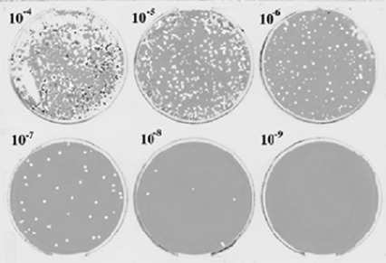
Formation of plaques
Virus count
- Counts only viable virus which are virus capable of multiplying.
- Must know the culture conditions for the virus studied.
- This method is useful for samples with very low virus count. (eg. food and water sample)
- Require time for incubation
Features
Virus count is a simple method which only works for viruses that infect monolayer cells and viruses that cause cell lysis. It is based on the principle that one virus on the monolayer produces one plaque.
4. Plants
Plant pathology also known as phytopathology is the scientific study of plant diseases caused by pathogens (infectious diseases) and environmental conditions (physiological factors). The use of plants to study virus is an expensive method. If viral growth occurs in the plant, there will be a reduce in quality and quantity of the crops. The virus plaques on the leaves determine the virus titers/numbers.
Tobacco Mosaic Virus (TMV)
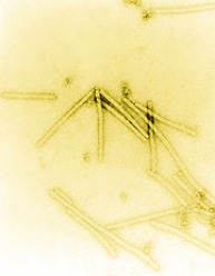
Electron micrograph of TMV particles
Tobacco mosaic virus (TMV) is an RNA virus that infects plants, especially tobacco and other members of the family Solanaceae. The infection causes characteristic patterns (mottling and discoloration) on the leaves.
Replication
Following entry into its host via mechanical inoculation, the Tobacco Mosaic Virus RNA genome is not immediately translated. Instead the RNA is processed by a mechanism that is not yet understood. The resulting mRNAs encode several proteins, including the coat protein and an RNA-dependent RNA polymerase (RdRp). Thus Tobacco Mosaic Virus can replicate its own genome. After the coat protein and RNA genome of Tobacco Mosaic Virus have been synthesized, they spontaneously assemble into complete Tobacco Mosaic Virus virions in a highly organized process. The protomers come together to form disks composed of two layers of protomers arranged in a helical spiral. The helical capsid grows by the addition of protomers to the end of the rod. As the rod lengthens, the RNA passes through a channel in its center and forms a loop at the growing end. In this way the RNA can easily fit as a spiral into the interior of the helical capsid.
Infection
When Tobacco Mosaic Virus infects a tobacco plant, the virus enters mechanically (through a ruptured plant cell wall) and replicates. After its multiplication, it enters the neighboring cells through plasmodesmata. For its smooth entry, Tobacco Mosaic Virus produces a 30 kDa movement protein called P30 which tends to enlarge the plasmodesmata. TMV most likely moves from cell-to-cell as a complex of the RNA, P30, and replicase proteins. The first symptom of this virus disease is a light green coloration between the veins of young leaves. This is followed quickly by the development of a “mosaic” or mottled pattern of light and dark green areas in the leaves. These symptoms develop quickly and are more pronounced on younger leaves. Mosaic does not result in plant death, but if infection occurs early in the season, plants are stunted. Lower leaves are subjected to “mosaic burn” especially during periods of hot and dry weather. In these cases, large dead areas develop in the leaves. This constitutes one of the most destructive phases of tobacco mosaic virus infection. Infected leaves may be crinkled, puckered, or elongated.
Cauliflower Mosaic Virus (CaMV)
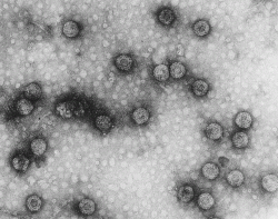
Cauliflower mosaic virus (CaMV) is the type member of the caulimoviruses, one of the six genera in the Caulimoviridae family, pararetroviruses that infect plants. Pararetroviruses replicate through reverse transcription just like retroviruses, but the viral particles contain DNA.
